
NeuraSense Labs Surgical sensing research
A new sense for surgery.
We develop impedance-based sensing and signal-analysis methods for surgical instruments, studying the electrical characteristics of tissue at the point of contact.
01 The question
Surgeons read tissue by sight and experience. Our research adds another perspective: the electrical characteristics of tissue.
Visual information is essential in surgery. Through a camera or a microscope, telling a nerve from a vessel, fat or connective tissue depends on appearance, anatomy and the surgeon’s judgment, and the view is not always clear.
Tissues also behave differently when a very small alternating current passes through them. That behavior is invisible, but it can be measured. Our research asks whether measurements taken through the instrument already in the surgeon’s hand can add a dependable second source of information, right where the instrument meets tissue.
02 From instrument to information
From tissue contact to electrical information.
Our development work follows four connected steps: contact, measurement, characterization and interpretation.
-
01 Contact
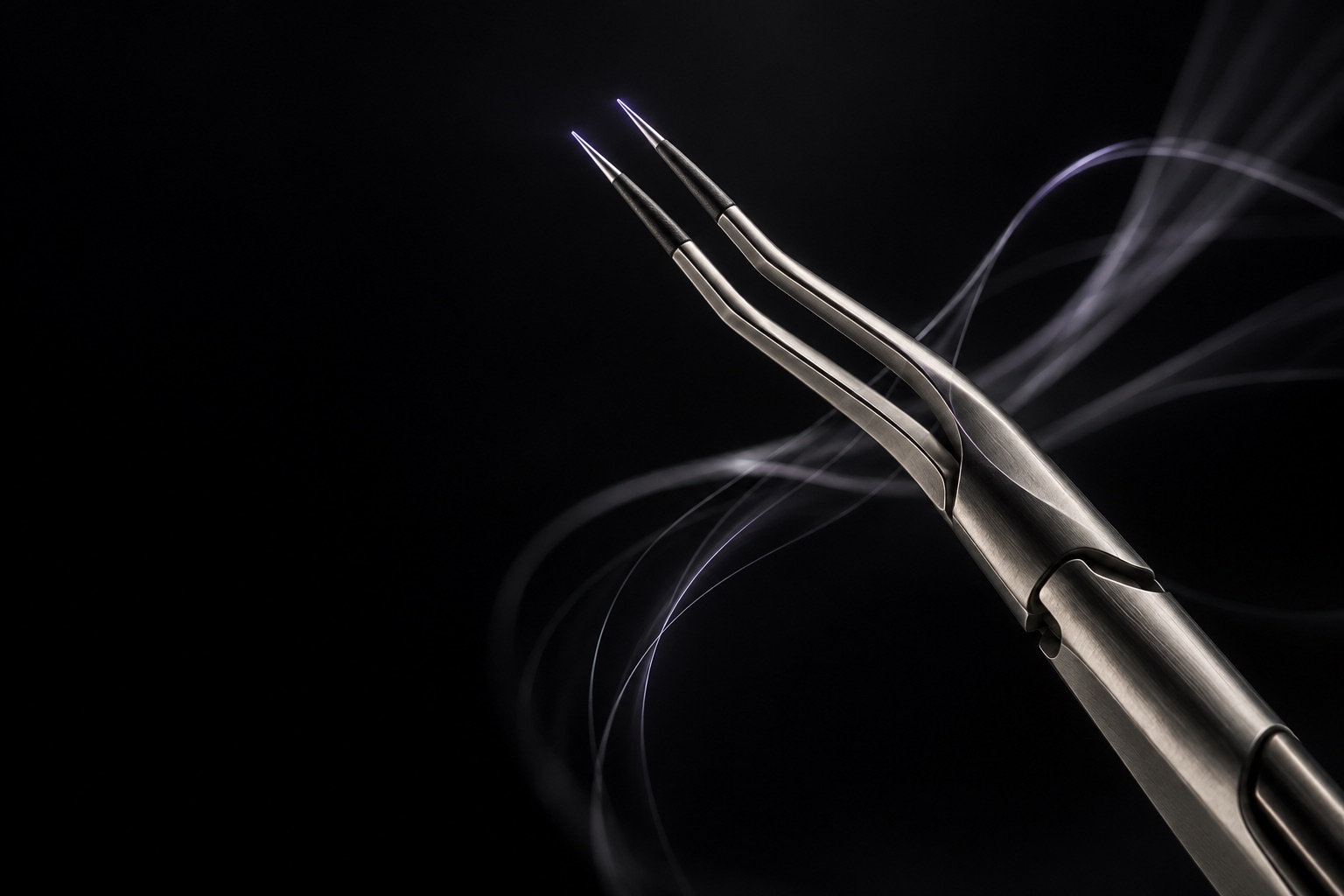
The sensing approach uses the tips of bipolar forceps as electrodes, placing the measurement at the instrument’s point of contact.
-
02 Measure
A small alternating test signal passes between the tips. The response is recorded two ways: how strongly the tissue opposes the current (magnitude) and how far the response shifts in time (phase).
-
03 Characterize
Repeating the measurement across many frequencies traces a spectrum. Cell membranes, fluid and fiber structure shape that spectrum, which is why different tissues can produce different patterns.
-
04 Interpret
Signal-analysis and classification methods compare spectra with characterized references. Our goal is useful tissue information that supports surgical judgment.
03 Research focus
Precision starts with measurement.
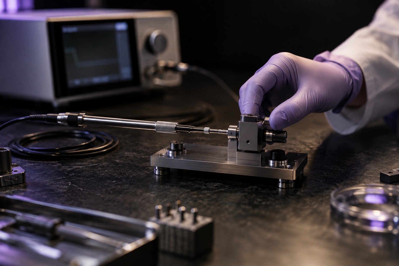
Reliable sensing starts with careful measurement, traceable data and tests that can be repeated. These are the priorities guiding our development work.
- A
Bench characterization
Measuring tissue impedance across frequencies under controlled conditions, to learn which differences are consistent and which are artifacts of the setup.
- B
Calibration and repeatability
Reference loads, drift checks and repeated measurements, so a signal means the same thing from one session to the next.
- C
Traceable data
Linking measurements to their specimens, setup and conditions, with separate data for model development and evaluation.
- D
Instrument integration
Carrying the measurement into a surgical instrument while respecting how that instrument is already held and used.

Insights
Short, educational explainers on the science and the method behind tissue sensing.
-
-
-
04 Where this leads
A path toward tissue-aware surgery.
Our roadmap starts with signal characterization and instrument integration, then extends toward tissue information for surgeons and future robotic systems.

-
Stage 1 · In development
Characterize the signal
Bench measurements of tissue impedance across frequencies, with calibration and repeatability built in from the start.
-
Stage 2 · In development
Bring sensing to the instrument
Developing an instrument-integrated measurement concept built around bipolar forceps, with bench and tissue-model evaluation as the next steps.
-
Stage 3 · Vision
Information at the point of contact
Clear tissue information at the point of contact that supports surgical judgment.
-
Stage 4 · Long-term vision
Tissue-aware surgical systems
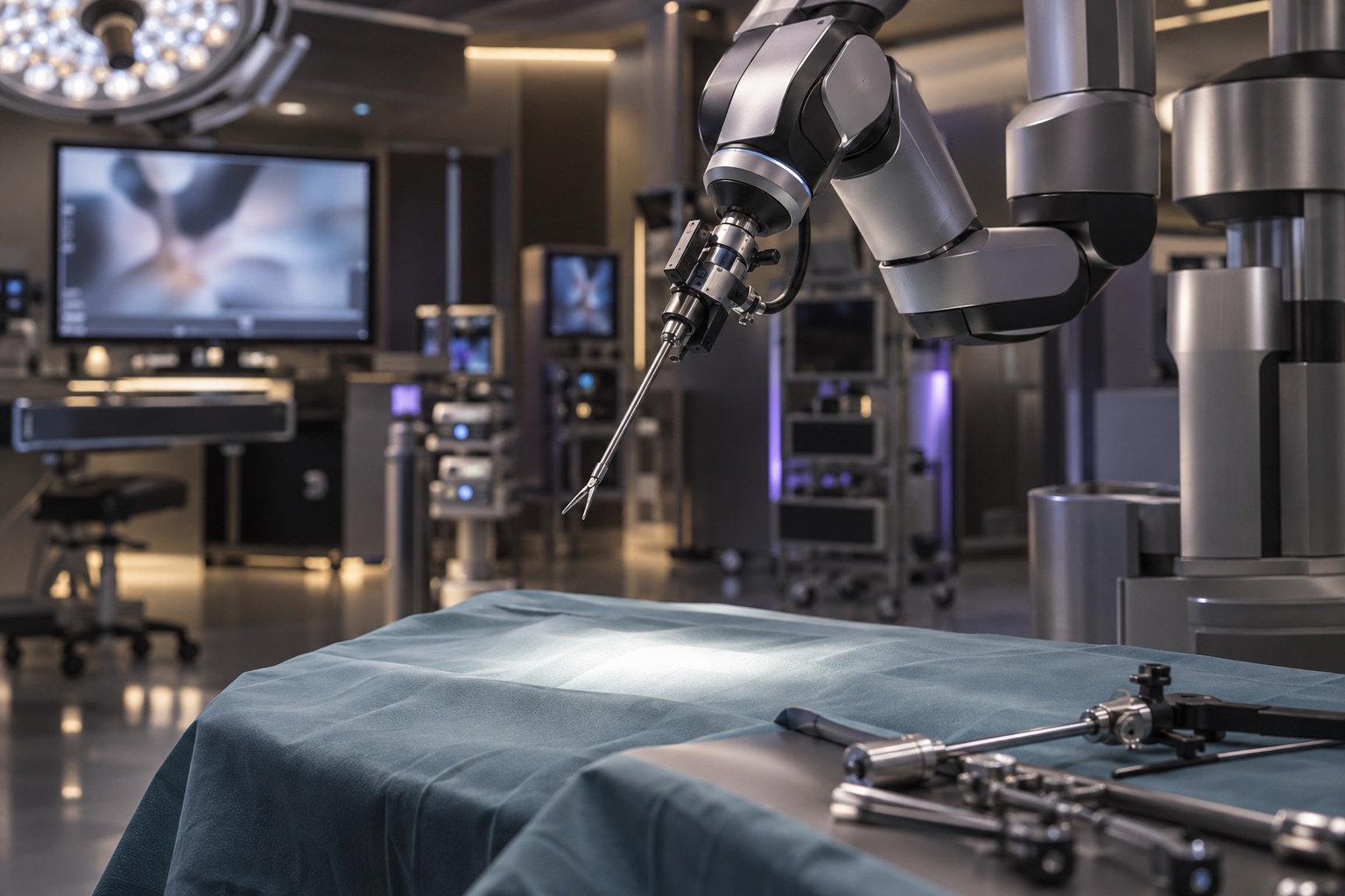
Tissue sensing as one input among imaging and instrument data in robotic and data-rich surgical environments.
05 Collaborate
Advance surgical sensing with us.
We welcome conversations with surgeons, researchers and engineers working on related problems in tissue sensing, instrumentation and surgical data.